 |
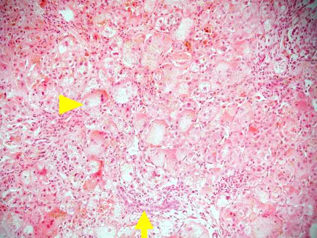
(Figure 1) Neonatal hepatitis with lobular disarray (arrowhead) and minimal mononuclear
infiltrate in the portal area (arrow). |
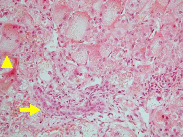 |
(Figure 2) Giant cell transformation of the hepatocytes (arrowhead) with increase in the
amount of the cytoplasm and multinucleation. In contrast to extrahepatic biliary atresia, no
proliferation of bile ducts, or edema and fibrosis around the bile duct is seen in the neonatal
hepatitis (arrow) |
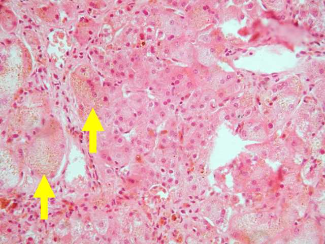 |
(Figure 3) Giant cell transformation of the hepatocytes (arrow) with increase in the
amount of the cytoplasm and multinucleation. Hepatic cholestasis with brown-green bile
pigments in the cytoplasm of the hepatocytes. |
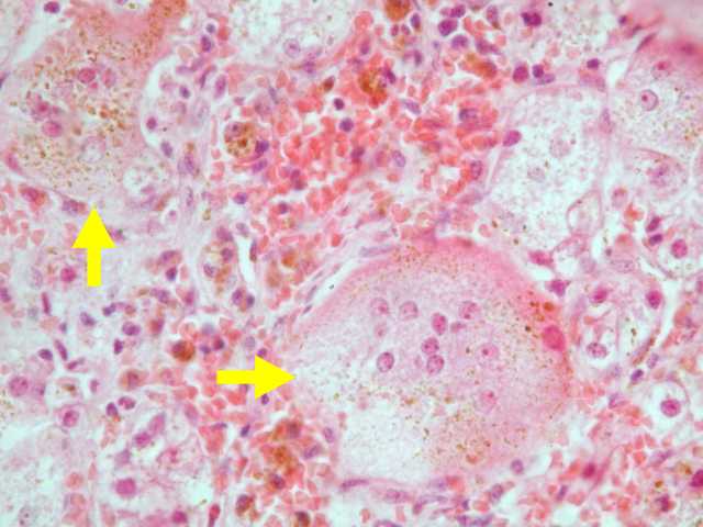 |
(Figure 4) Giant cell transformation of the hepatocytes (arrow) with increase in the
amount of the cytoplasm and multinucleation. Hepatic cholestasis with brown-green bile
pigments in the cytoplasm of the hepatocytes. |
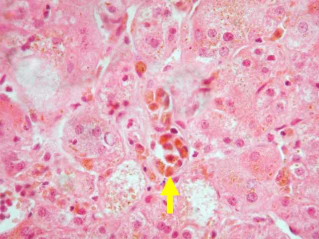 |
(Figure 5) Kuppfer cell proliferation with bile pigments in their cytoplasm (arrow). |