 |
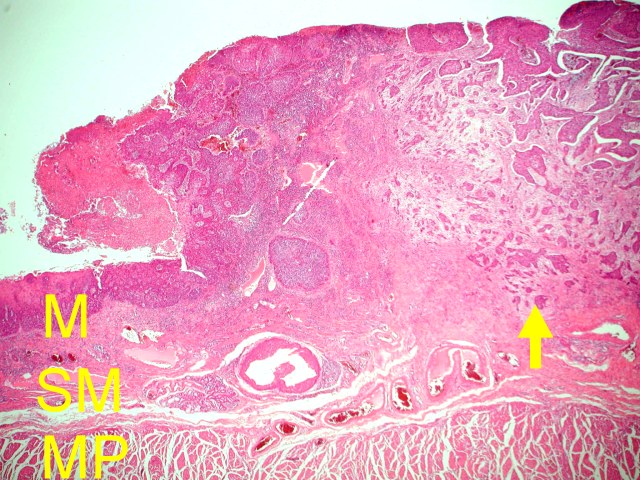
Figure 1) Squamous cell carcinoma of esophagus (arrow). The carcinoma
has invaded the submucosa.(M:mucosa, SM: submucosa, MO: muscularis propria). |
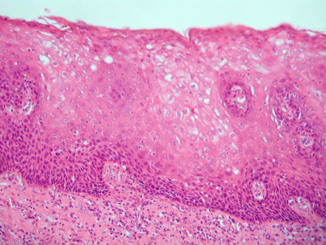 |
(Figure 2) Normal squamous epithelium of esophagus. Note the
orderly maturation of the epithelium with gradually increase in
the amount of cytoplasm and decrease of the nucleus to cytoplasm
ratio. |
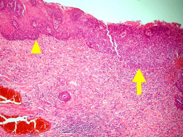 |
Figure 3) Carcinoma in situ of the squamous epithelium (arrow).
Carcinoma in situ lesion is seen adjacent to the invasive carcinoma
(not shown here). The basement membrane is intact in carcinoma in
situ areas. Normal epithelium is seen (arrowhead) |
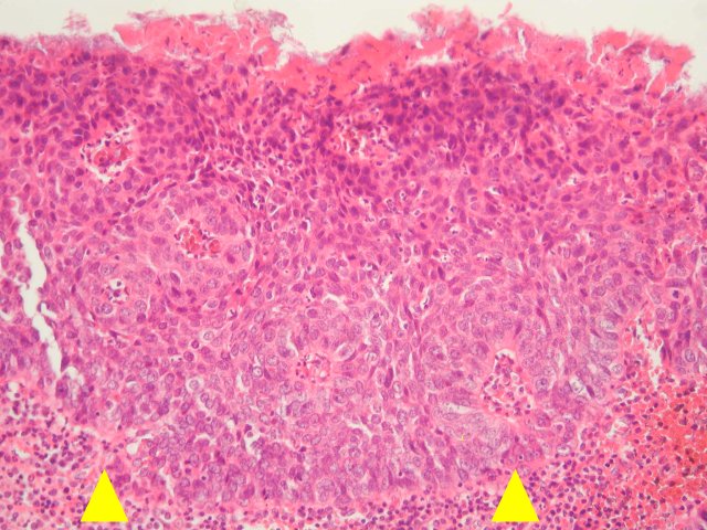 |
(Figure 4)Carcinoma in situ of squamous epithelium. The whole layers
of the epithelium is replaced by dysplastic cells with enlarged and
hyperchromatic nuclei. The basement membrane is intact (arrowhead). |
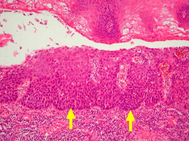 |
(Figure 5)Carcinoma in situ of squamous epithelium (arrow). The
|
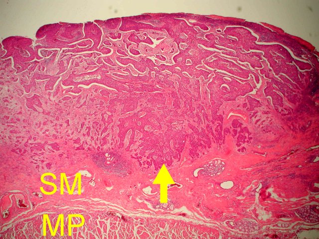 |
(Figure 6) Invasive squamous cell carcinoma with irregular infiltrating
borders (arrow). The carcinoma has invaded the submucosa layer. |
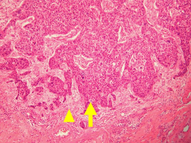 |
(Figure 7) Invasive squamous cell carcinoma with irregular infiltrating
borders (arrow). Desmoplastic stroma is seen. (arrowhead) |
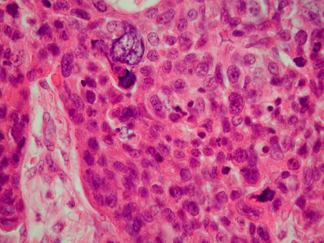 |
Figure 8) Mark nuclear pleomorphism and hyperchromasia in
carcinoma cells. Note high nucleus to cytoplasm ratio
irregular nuclear membrane, and frequent mitoses. |
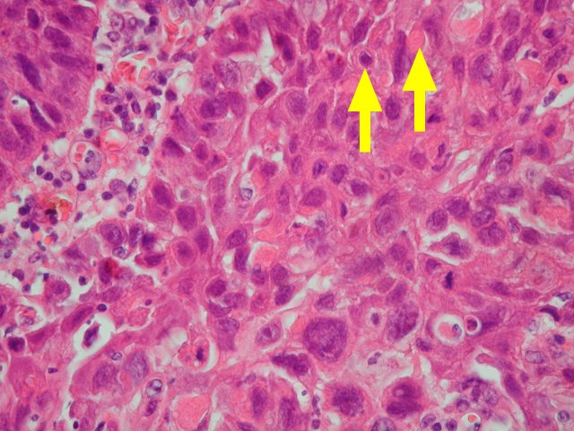 |
(Figure 9) Focal cancer cell keratinization (arrow) |