 |
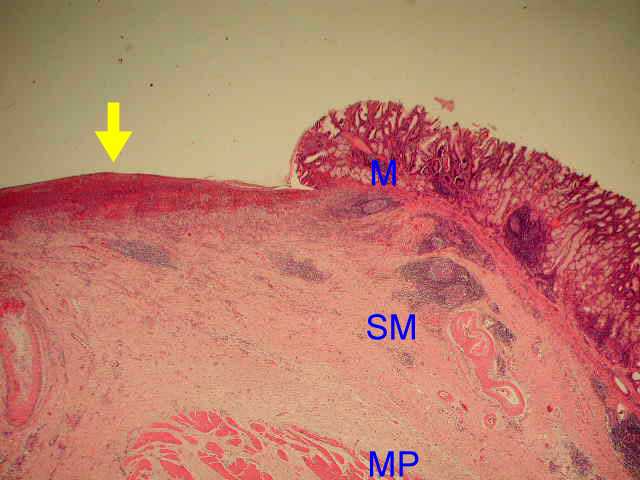
(Figure 1) Peptic ulcer of stomach (arrow). The whole mucosa and part of
submucosa are denuded.(M: mucosa, SM: submucosa, MP: muscularis propria) |
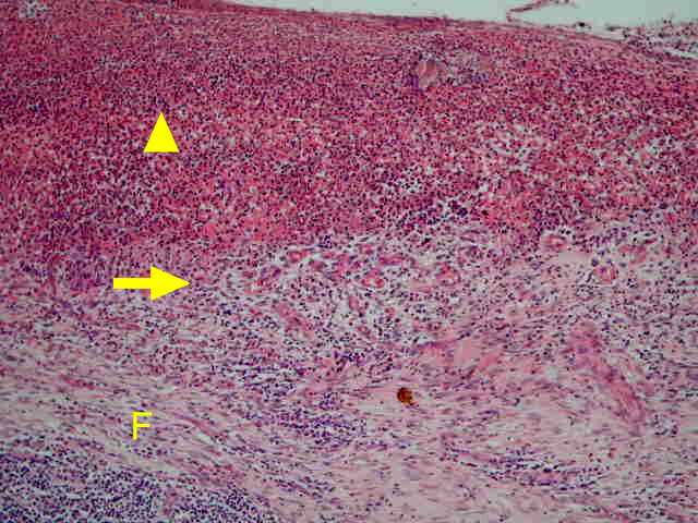 |
(FIgure 2). Four zones of active peptic ulcer.The necrotic fibrinoid debris and
nonspecific inflammatory infiltrate are labeled by arrowhead. Beneath the
necrotic and inflammatory zones, there is granulation tissue (arrow). Below
the granulation tissue, fibrotic tissue is seen (F). |
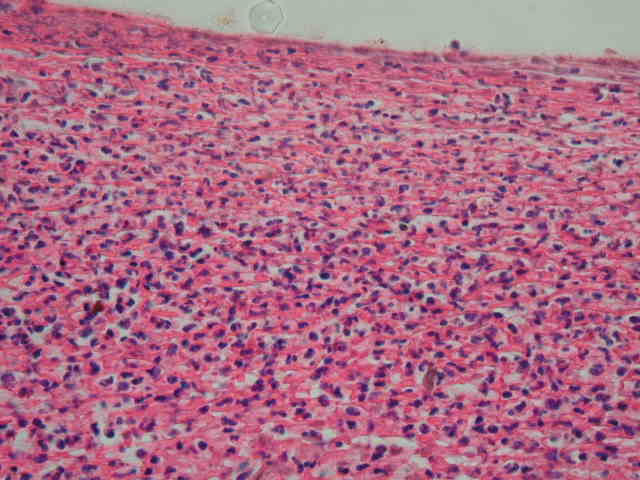 |
(Figure 3). Necrotic fibrinoid debris and inflammatory infiltrate in the ulcer
base. |
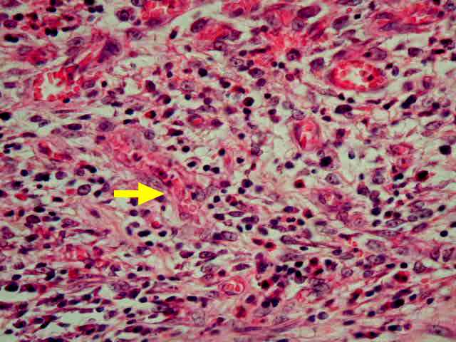 |
(Figure 4). Granulation tissue in the ulcer base. New blood vessels lined by
plump endothelial cells (arrow). Edema and inflammatory infiltrate are also seen. |
 |
(Figure 5). Fibrotic tissue beneath the ulcer base. |
 |
(Figure 6). Fibrosis in the muscularis propria (arrow). Chronic inflammatory
cell infiltration is also noted (arrowhead).
|
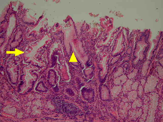 |
(Figure 7) Chronic gastritis with intestinal metaplasia (arrow) seen in the
mucosa around the peptic ulcer.The mucinous gastric foveolar epithelium
(arrowhead) is replaced by intestinal type of epithelium (arrow). |
 |
(Figure 8) Intestinal metaplasia in chronic gastritis. The gastric foveolar
epithelium (arrowhead) is replaced by intestinal type of epithelium (arrow).
The intestinal epithelium has goblet cells. Chronic inflammatory cell
infiltration is also seen. |